
Equipment
Leica Stellaris 8 & 5 Confocal Microscopes
High-end spectral confocal systems with a white light laser (440–790 nm tunable excitation) and advanced HyD detectors for photon counting. The Stellaris 8 includes fast resonant scanning and fluorescence lifetime imaging (FALCON FLIM) capabilities, allowing high-speed live-cell imaging and functional imaging of fluorescence lifetimes. Both systems feature Leica’s TauSense and LIGHTNING super-resolution processing for enhanced resolution and image quality.
The STELLARIS 8 confocal microscope from Leica Microsystems integrates a multitude of advanced features:
- Power HyD Detectors: The Power HyD S detectors offer analog and photon counting capabilities for confocal imaging. The Power HyD R detectors are optimized for near-infrared imaging, and the Power HyD X detectors are specialized for FLIM and FCS. ref
- White Light Laser (WLL): This system allows for the use of up to 8 independent laser lines for simultaneous scanning. Users can precisely tune these excitation lines within a modified range of 440 nm – 790 nm to match the spectral characteristics of the dyes in their samples, ensuring optimal excitation of each fluorophore. Next to the WLL, the system has a solid state 405 and 488 laser. ref
- 8Khz Resonant Scanner: The STELLARIS platform can uses an 8 kHz resonant scanner to maintain high-speed image acquisition, allowing detailed observation of dynamic processes at the nanoscale.
- TauSense Technology: This suite of imaging tools is based on fluorescence lifetime and enhances confocal experiments by providing functional information, improving image quality, and enabling the separation of fluorescent signals with similar spectra. ref
- FLIM and FCS: The STELLARIS 8 FALCON FLIM offers functional imaging using fluorescence lifetime, enabling researchers to investigate cellular physiology and explore dynamics in living cells. It allows for rapid kinetic studies and adds new dimensions of contrast for biosensing and protein interactions. ref ref
- LIGHTNING Detection Concept: Offers adaptive image reconstruction in real-time and 5D, resolving nanoscale structures down to 120 nm and supporting simultaneous multicolor super-resolution imaging. It fully integrates into the online image acquisition process for optimal information extraction. ref
- OKO lab Incubator: Compatible with all STELLARIS configurations, this incubator controls temperature around the microscope with a range from 3°C above ambient to 45°C, and regulates CO2 between 0 and 30%, ensuring stable and accurate conditions for live-cell imaging
Help and tutorials:
Stellaris 8 (and 5) help This the complete helpfile of the Stellaris LAS-X (only available from AMC or CDW)
Leica YouTube tutorials
Leica Training tutorials (beta) (password protected, ask us for the password)

Leica Stellaris Spectraplex Confocal Microscope
Leica STELLARIS SpectraPlex is a high-multiplex spatial biology solution integrated into a confocal platform. It is specifically engineered to overcome the limits of traditional multicolor imaging by allowing researchers to visualize 15+ markers simultaneously in a single round of staining and imaging. Using a White Light Laser (WLL) and advanced spectral unmixing algorithms, SpectraPlex captures 3D spatial information with subcellular precision while minimizing crosstalk. The system is designed for complex tissue analysis—such as cancer immunology and neuroscience—and features a “Virtual Fridge” for digital panel design and seamless integration with Aivia AI for automated cell phenotyping and clustering.

Leica Cell DIVE
The Leica Cell DIVE is a high-content multiplexed imaging platform designed to visualize and analyze over 60 biomarkers on a single tissue section. By utilizing an automated, robotic, iterative cycle of staining, imaging, and dye inactivation, the system generates high-dimensional spatial data while preserving tissue integrity for downstream analysis.
Unlike closed platforms restricted to proprietary panels, Cell DIVE operates as an open system. This allows for the utilization of primary antibodies from any source alongside a library of over 350 validated markers. This flexibility, combined with whole-slide imaging capabilities, enables comprehensive phenotyping and spatial analysis of complex tissue microenvironments at true single-cell resolution. It is optimized for spatially resolved proteomics, immuno-oncology, and extensive biomarker discovery in both FFPE and frozen tissues.

Leica LMD-7
The Leica LMD-7 is a high-performance laser microdissection system designed for the precise isolation of specific single cells, distinct cell clusters, or tissue regions from heterogeneous samples. This platform enables researchers to obtain pure, contamination-free starting material necessary for sensitive downstream molecular analysis, including Next-Generation Sequencing (NGS), transcriptomics, and proteomics.
Distinguished by its unique gravity-based collection method, the LMD-7 cuts samples using a high-powered, steerable laser, allowing the dissectate to fall directly into a capture vessel without mechanical contact or the use of contamination-prone adhesives. This contact-free process ensures high sample integrity and allows for the use of standard consumables. The system features adjustable high-power laser optics capable of cutting through a wide variety of specimens, from thin tissue sections and smears to hard plant material and bone, making it a versatile tool for molecular pathology and neurobiology

Leica Mica Microscope
An integrated wide-field + confocal fluorescence microscope with built-in incubator. The Leica Mica “Microhub” allows researchers to seamlessly switch between multiple imaging modes (widefield, confocal, Z-stacks, time-lapse, etc.) within one enclosed system. Users can quickly capture a large field-of-view overview at low magnification, then zoom in and activate confocal mode on regions of interest without moving the sample. Mica also features on-board computational clearing (Thunder) and Lightning deconvolution for real-time removal of out-of-focus blur, as well as full environmental control (temperature, CO₂, humidity) to keep live cells in physiological conditions during imaging.
Microscope
Mica unites widefield and confocal imaging in a sample protecting, incubating environment. With the simple push of a button, you have everything you need – all in one place – to supercharge fluorescence imaging workflows and get meaningful scientific results faster.
Select the right modality in real time
Mica unifies transmitted and fluorescence light imaging modalities. You can select from multiple imaging modalities all within one Microhub, including widefield, confocal, THUNDER imaging, LIGHTNING, Z-stacks, time-lapse and more.
This enables you to
- generate fast overviews with widefield at low magnification
- gradually zoom in on the regions of interest
- switch to confocal when and where needed without ever moving the sample to a different system
Video Tutorials
Mica Videos
Mica Videos Tutorials on Youtube
Leica YouTube tutorials
Leica Training tutorials (beta) (password protected, ask us for the password)
Online Help
Mica Help (only available from AMC or CDW)
Achieve physiological-like conditions thoughout your experiments
Live cell experiments require the cells to be in optimal shape. Typically, 2D and 3D cells in media require the temperature and the pH (via CO2) in the environment to be controlled. Stable nutrition and ion concentrations require the evaporation to be minimal. Some experiments even demand the O2 to be mimicked closer to physiological levels. Mica can provide the right conditions in the live cell configuration.
- Mica is an incubator: the entire encapsulated inner sample space can be climate controlled (temperature, CO2 and humidity regulation) and offers ideal conditions for short and long-term live cell observation.
- From dark to light: Mica also enables you to enjoy a brightly lit lab—freeing you from the constraints of sitting in a dark room for hours monitoring your experiment.
Radically simplified workflows
Intelligent automation and AI-supported analysis enables greater efficiency and a faster track to publication.
- Reduce over 60% of process steps through system intelligence
- Reduce time and effort from sample to insight by simplifying your entire workflow
- Enable 100% reproducibility and repeatability throughout your experiment
Objectives:
- N PLAN 2.5 x / 0.12 Dry (used for overviews of sample)
- HC Plan Apo 10 x / 0.40 CS2 Dry
- HC Plan Apo 20 x / 0.75 CS2 Dry
- HC PL APO 63 x / 1.20 CS2 Water (motCorr), (Automatic water pump)
Thunder Algorithms
3 dedicated algorithms are available that remove out-of-focus light in real-time, thus providing fast optical sectioning capabilities:
- Instant Computational Clearing (ICC): Removal of background and out-of-focus light.
- Small Volume Computational Clearing (SVCC): ICC and deconvolution algorithm for thin samples (e.g. cells, yeast)
- Large Volume Computational clearing (LVCC): ICC and deconvolution algorithm for thick samples (e.g. tissue sections, embryos, organoids).
The software used is Leica LAS-X

Leica Thunder Widefield Microscope
An inverted fluorescence microscope equipped with real-time computational clearing. The Thunder provides high-contrast wide-field images by removing out-of-focus light on the fly, approaching confocal-like sectioning in thick samples. It is ideal for fast imaging of fluorescent samples where gentle acquisition and quick results are needed (e.g. screening or live-cell snapshots).
Microscope
Some info: THUNDER Revealing Image Detail Hidden in the Haze , Overview PDF , Spheroid-Imaging The system is built on a fully automated, inverted Leica DMI8 microscope equipped with AFC (hardware autofocus control) and the objectives below.
Objectives:
- N PLAN 5 x / 0.12 Dry
- HC PL Fluotar 10 x / 0.32 Dry
- HC PL Fluotar 20 x / 0.40 Dry
- HC PL Fluotar 40 x / 0.60 Dry
- HC PL APO 63 x / 1.40 Oil
Fluorescence Filters
| Name | LED | Excitation filter | Dichroic mirror | Emission filter |
| CFP | 440 | BP 436/10 | 455 | BP 480/40 |
LED Excitation, Quad Filter Block (Dichroics/Filters) and Filter Wheel
Two multiband filter cubes are available (see below). The first is a quad filter set that uses an external filter-wheel to avoid crosstalk between different dyes. The second is a standard multiband filter cube for cyan, yellow, red and near infrared emission dyes).
| Quad Filter Block | LED | Excitation Range | Dichroic | Emission Range | Emission Filter wheel | FLUOROPHORES |
| DFT51010 | 390 | 375-407 | 415 | 420-450 | BP440/40 | DAPI, Hoechst |
| 475 | 462-496 | 490 | 506-532 | BP510/40 | FITC, GFP, Alexa488 | |
| 555 | 542-566 | 572 | 578-610 | BP590/50 | Alexa568, Rhodamine, DsRed-2 | |
| 635 | 622-654 | 660 | 666-724 | BP705/72 | Alexa647, Cy5 | |
| CYR70010 | 440 | 422-450 | 459 | 462-484 | 100 % | CyanFP |
| 510 | 495-517 | 523 | 527-551 | 100 % | YellowFP | |
| 575 | 566-590 | 598 | 602-680 | 100 % | mCherry, Alexa594, Texas-Red | |
| 730 | 710-750 | 763 | 770-850 | 100 % | nIR Autofluorescence |
Motorized XY Stage ITK QUANTUM
For fast and precise sample positioning and mosaic/multiposition acquisitions (see specifications below). It is provided with several inserts to hold slides, plates and 96-multiwell plates.
- Travel range 122 x 82mmSpeed 500mm/sec
- Resolution 5 nm
- Reproducibility < 1 μm
- Accuracy < +/- 1 μm
Leica DFC9000 GTC sCMOS camera
- 4.2 MPixel
- Image sensor type: Front-illuminated scientific CMOS CIS2020A
- Shutter: Rolling shutter with global reset
- Pixel: chip of 2048x2048 pixels; 6.5 mm pixel size
- Speed of Acquisition: 50 fps (USB 3.0); 90 fps (Camera Link)
- Maximum quantum efficiency: 82% @580 nm
- Binning: 2x2, 3x3, 4x4, 8x8
- 12 bit and 16 bit digitalizers
Environmental control
UNO Stage-Top Incubator (OKO-LAB). Includes #H301-K stage incubator equipped with inserts for 2 petri dishes and 1 slide, universal multi-well plates, 12-well plates (and more). CO2 and humidity are regulated through the manual UNO-H-T-CO2 controller (OKO-LAB) equipped with an Air/CO2mixer, an air pump and a humidity module.
Thunder Algorithms
3 dedicated algorithms are available that remove out-of-focus light in real-time, thus providing fast optical sectioning capabilities:
- Instant Computational Clearing (ICC): Removal of background and out-of-focus light.
- Small Volume Computational Clearing (SVCC): ICC and deconvolution algorithm for thin samples (e.g. cells, yeast)
- Large Volume Computational clearing (LVCC): ICC and deconvolution algorithm for thick samples (e.g. tissue sections, embryos, organoids).
The software used is Leica LAS-X

ZEISS Axioscan 7
The ZEISS Axioscan 7 is a high-performance, fully automated slide scanner designed for high-throughput digitization in life sciences and geology. It stands out for its versatility, allowing users to switch rapidly between brightfield, fluorescence, and polarization imaging modes while accommodating up to 100 slides in a single run. The system features advanced hardware like the Colibri 7 LED light source and high-speed filter wheels, which enable gentle yet rapid multispectral fluorescence imaging of up to nine channels.
Capacity: Loads up to 100 standard slides (26 mm x 76 mm). Speed: Fast “flash mode” for brightfield and motorized switching for complex multiplexing. Imaging Modes: Brightfield, Fluorescence, Polarization (Circular/Linear), and TIE (Phase/Relief) contrast. Precision: Features the Axiocam 705 (color) and Axiocam 712 (monochrome) for research-grade resolution.

Leica DM6 B Upright Microscope
A versatile wide-field microscope for slides and fixed samples. The Leica DM6 supports brightfield, phase contrast, and fluorescence imaging modes. It is equipped with both a monochrome camera (for capturing fluorescently labeled samples) and a color camera (for histology or colorimetric stains), allowing users to document samples in multiple modalities. This system is often used for imaging tissue sections, histology slides, and other specimens mounted on glass slides.

Zeiss Axio Zoom.V16 Macro Fluorescence Microscope
A fluorescence macro zoom microscope designed for imaging large or whole-mount samples. The Axio Zoom.V16 provides a high zoom range with both fluorescence and brightfield illumination options. An optional ApoTome optical sectioning device allows for structured illumination to obtain optically sectioned images of fluorescent samples (reducing background in thicker specimens). This instrument is useful for capturing overview images of large specimens (e.g. embryos, organoids, small animals) with the ability to optically slice in Z for 3D information.

Leica DMi8 Live-Cell Imaging System
An inverted research microscope with an environmental chamber for live-cell imaging. The DMi8 is configured for long-term time-lapse experiments, featuring hardware autofocus and an incubator enclosure to maintain stable temperature, CO₂, and humidity for cultured cells. This system allows researchers to observe dynamic cellular processes over hours or days under controlled conditions (e.g. studies of cell migration, division, or drug responses in real time).

Molecular Devices ImageXpress Pico (High-Content Imager)
An automated digital microscope for high-content screening. The ImageXpress Pico can run fully automated brightfield and multi-wavelength fluorescence imaging of multi-well plates. It includes a library of preconfigured protocols for common cell-based assays and on-the-fly image analysis capabilities. Features such as digital confocal deconvolution, live preview scanning, and automated cell segmentation enable users to quickly obtain quantitative data from fluorescence or brightfield assays. With its built-in incubation option, long-term live-cell assays in multiwell plates can be performed, making it a powerful tool for drug screening and phenotypic assays.

IncuCyte S3 Live-Cell Analyzer
A real-time live-cell imaging and analysis platform housed inside a standard incubator. The IncuCyte S3 automatically acquires phase-contrast and fluorescence images of cell cultures at defined intervals, allowing kinetic measurements of cell behavior (proliferation, morphology changes, fluorescence expression, etc.) over days or weeks. Because the imaging occurs in an incubation environment, cells remain undisturbed and in optimal conditions throughout the experiment. The system’s software provides quantitative metrics in real time, enabling time-lapse studies of living cells with minimal intervention.

Leica CryoStat CryoStar NX70
A cryostat instrument for frozen tissue sectioning (sample preparation). This unit allows researchers to prepare ultrathin frozen sections of tissues or cell pellets at temperatures around -20°C to -30°C. Cryosectioning is often used to preserve antigenicity for immunofluorescence or to prepare samples for ultrastructural analysis. The cryostat is available to trained users for quick sample freezing and sectioning on-site.

General information
Talos L120c
A high-end Thermo Fisher Scientific 120kV Transmission Electron Microscope (TEM) equipped with a high-resolution bottom mount camera (BM-Ceta 16M); this combination makes for high resolution images at high magnifications. To fully utilize these specs, this microscope also has software for mapping, tomography (with tomography holder) and embedded in the imaging software, STEM mode can also be selected. Another high-end feature to this microscope is the EDS detector (Bruker Quantax 200 XFlash 6T detector), that can do elemental analyses on whole regions altogether (TEM-EDS) or more specific while showing the localization of elements (STEM-EDS).

Tecnai G2 Spirit Biotwin
This work-horse has earned it’s stripes by being in the EM center for over 10 years already! A FEI (now Thermo Fisher Scientific) 120kV Transmission Electron Microscope (TEM) aligned at 80kV to increase stability and contrast. Equipped with two EMSIS camera’s: a Xarosa (20MP CMOS) bottom mount and a Veleta side-mount.

JEM-1400 Flash
The newest addition to the EM center: a Jeol 120kV Transmission Electron Microscope (TEM). Aligned at both 80kV and 120kV for easy switching between high contrast and high-resolution imaging using the bottom mount EMSIS Xarosa (20MP CMOS) camera. Besides a standard Quick Change holder, a Quartet Multi-grid holder is also available to enable high throughput imaging by loading up to four samples at once (for magnifications up to 40.000x).

Gemini Sigma 300
Except for a new PC and updated software version, this Zeiss Field Emission Gun Scanning Electron Microscope (FEG-SEM) has also been in the EM center for over 10 years already! Equipped with two secondary electron detectors (SE2 and InLens) and a primary/backscatter electron detector (HDBSE), there is a variety of ways to image samples, as both one or two signals can be used for the image at once. Samples are typically loaded using our 9-spot carousel holder, but other holders (e.g. for fixed tilt) are also available.

Ultramicrotomy Equipment
Proper sample preparation is critical for electron microscopy, and the facility provides ultramicrotomes for sectioning samples. We have a Leica Ultracut UC7 ultramicrotome for room-temperature sectioning of resin-embedded samples, as well as a Leica Cryo-Ultramicroscope for cutting ultrathin frozen sections (cryo-sections). These instruments allow users to cut sections ~50–100 nm thin for TEM grids or slightly thicker sections for array tomography. The usage of ultramicrotomes is available to trained users at a nominal hourly rate, or our EM staff can perform sectioning as a service (especially for difficult samples like tissues that need optimal sectioning technique). We also have equipment for routine EM sample prep, such as resin embedding kits, a sputter coater for SEM sample gold coating, and critical-point dryer for fully dehydrating samples, ensuring comprehensive preparation capabilities.
Correlative Light-Electron Microscopy (CLEM) Setup
To facilitate CLEM projects, the EM facility can coordinate the use of fluorescence microscopes alongside EM. We provide support for marking regions of interest in fluorescently labeled samples (for example, using a fluorescent wide-field microscope to identify cells of interest on a coverslip or section, then correlating those coordinates in the electron microscope). We have specialized tools like finder grids, reference markers, and software to overlay fluorescence images with EM images. This allows researchers to pinpoint specific targets (e.g. a fluorescent protein-tagged structure) in the EM for high-resolution visualization. Our team has experience with immuno-labeling for EM (using gold-conjugated antibodies) and subsequent correlation with fluorescence, enabling powerful multimodal imaging of the same specimen.
Analyzers
BD FACSCanto II
The FACSCanto II is a reliable, easy-to-learn flow cytometer equipped with three lasers (405 nm, 488 nm, and 633 nm), allowing detection of up to eight fluorescent markers in a single sample. The instrument also includes an HTS (High-Throughput Sampler) for running samples directly from multi-well plates, supporting higher-throughput workflows. Overall, the FACSCanto II is a user-friendly workhorse, suitable for most standard immunophenotyping experiments, and is accessible to new users after training.

BD Symphony A1
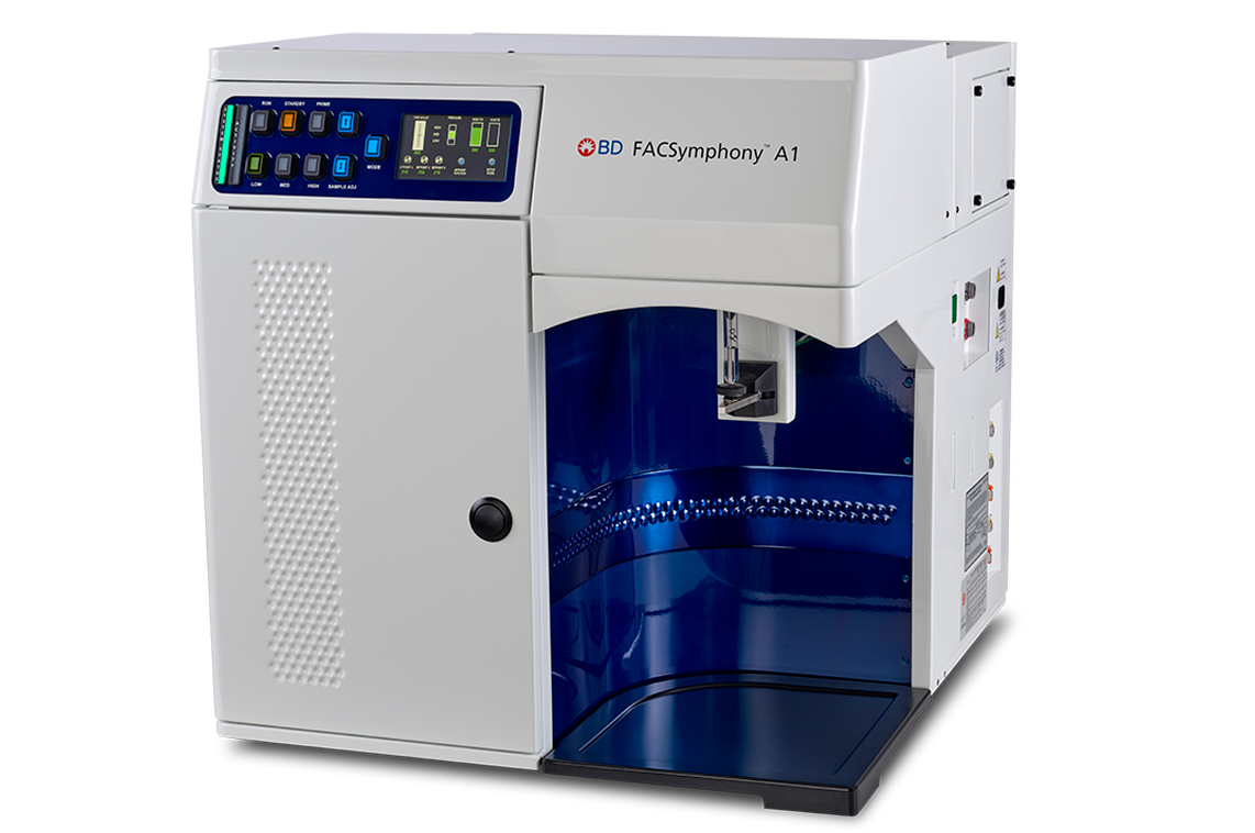
The Symphony A1 is a high-parameter flow cytometer with four lasers (405 nm, 488 nm, 561 nm, and 637 nm), enabling detection of up to 16 fluorescent markers per sample. It also includes an HTS (High-Throughput Sampler) for convenient plate-based acquisition. This system is ideal for deep immunophenotyping or high-dimensional single-cell studies, while remaining approachable to new users after training.
Sony ID7000 Spectral Cytometer
The Sony ID7000 is a high-performance spectral flow cytometer with six lasers (320 nm, 355 nm, 405 nm, 488 nm, 561 nm, and 637 nm), providing broad excitation for many fluorophores. Its spectral detection and unmixing capabilities allow accurate resolution of overlapping emissions, supporting large, high-dimensional multi-color panels for advanced immunophenotyping and single-cell analysis. The ID7000 also supports plate-based sampling for high-throughput or automated workflows. It is ideal for deep immune profiling and spectral panel development, and is accessible to new users with training.
Cytek Amnis ImageStream X Mk II
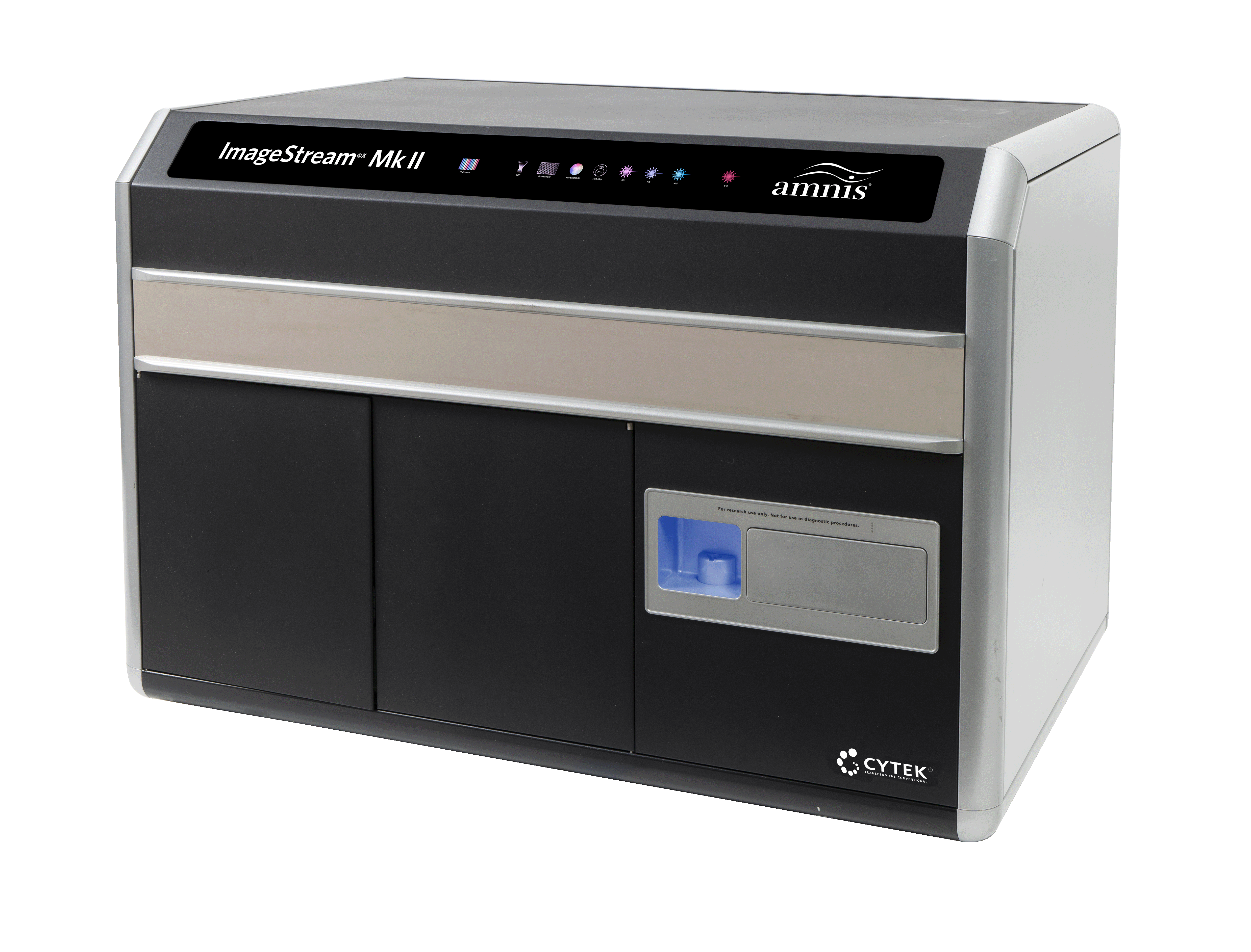
The ImageStream X Mk II combines flow cytometry with high-resolution microscopy. It has four lasers (405 nm, 488 nm, 561 nm, and 642 nm) and can measure up to 10 fluorescent parameters, while capturing brightfield and fluorescent images of every cell. This allows analysis of cellular features such as nuclear translocation, cell conjugates, and morphology, which cannot be discerned from scatter plots alone. The system uses objectives up to 60×, providing high-resolution imaging (~0.1 µm/pixel), and includes a plate loader for high-throughput experiments. The ImageStream X Mk II is ideal for correlating cellular phenotype with spatial distribution of signals, combining the quantitative power of flow cytometry with detailed imaging, while remaining accessible to trained new users.
Sorters
Sony FP7000
The Sony FP7000 is a high-parameter spectral cell sorter equipped with up to six lasers (320 nm, 349 nm, 405 nm, 488 nm, 561 nm, 637 nm) and 182 detectors, enabling analysis and sorting of panels with more than 40 colors.
Its real-time spectral unmixing system allows separation of overlapping fluorescence signatures and treats autofluorescence as a separate parameter, improving accuracy in complex, highly multiplexed experiments.
The FP7000 supports multi-way sorting, multiple collection formats (tubes or plates), and various nozzle sizes (70 µm, 100 µm, 130 µm) to optimize for different cell types and sizes. Guided workflows and automated setup make it usable for both new and experienced users.

Sony SH800 Cell Sorter
The Sony SH800 is a benchtop fluorescence-activated cell sorter (FACS) with four colinear lasers (405 nm, 488 nm, 561 nm, and 638 nm) and detection of up to six fluorescent markers. It can perform two-way sorting, depositing two different populations simultaneously, and supports multiple collection formats, including 15 mL and 5 mL tubes, Eppendorf tubes, and multi-well plates, with single-cell sorting into 96- or 384-well plates. The SH800 also features index sorting for clonal tracking. Multiple nozzle sizes (70 µm, 100 µm, 130 µm) allow sorting of different cell types at optimal speed and purity. Overall, the SH800 is easy to use, versatile, and suitable for a wide range of cell sorting applications, making it accessible to both new and experienced users.

BD Symphony S6
The BD Symphony S6 is an advanced fluorescence-activated cell sorter (FACS) with four lasers (405 nm, 488 nm, 561 nm, and 640 nm) and detection of up to 16 fluorescent markers. It can perform six-way sorting, allowing up to six populations to be purified simultaneously into separate collection tubes. It supports a variety of collection formats including tubes, multi-well plates, and slides, and offers single-cell index sorting. Multiple nozzle sizes (70 µm, 85 µm, 100 µm, 130 µm) allow sorting of cells of different sizes at optimal speed and purity.

All workstations are in Room M3-119 unless marked Remote-only. Use the filter to find stations or software.
On-site Workstations
| Station | Software Installed |
|---|---|
| Microscopy A | Leica LAS-X (+2D/3D Analysis + FLIM), ImagePro Plus 7.0, Zeiss ZEN 2012, Aivia (2D/3D), ImageJ, ImageScope, IncuCyte S3 |
| Microscopy B | Leica LAS-X (+2D Analysis), SymPhoTime 64, ImagePro Plus 7.0, ImageJ, ImageScope, IncuCyte S3 |
| Microscopy C | Leica LAS-X (+2D/3D Analysis), Imaris (2D/3D), ImageJ, ImageScope, IncuCyte S3 |
| Microscopy D | Leica LAS-X, Huygens Pro (2D/3D deconvolution), ImageJ, ImageScope, IncuCyte S3 |
| Flow A | FlowJo 10, FCS Express 7, Cytosplore, Leica LAS-X Core, ImageJ, ImageScope, IncuCyte S3 |
| Flow B | FlowJo 10, FCS Express 7, Cytosplore, Leica LAS-X Core, ImageJ, ImageScope, IncuCyte S3 |
| Flow C | FlowJo 10, FCS Express 7, Sony ID7000 software, Cytosplore, Leica LAS-X Core, ImageJ, ImageScope, IncuCyte S3 |
| Flow D | FlowJo 10, FCS Express 7, Cytosplore, Leica LAS-X Core, ImageJ, ImageScope, IncuCyte S3 |
Remote-only Workstations Remote-only
Accessible via CDW/OneView or AMC PC
| Station | Software Installed |
|---|---|
| Mix A | FlowJo 10, FCS Express 7, Cytosplore, CellProfiler, Leica LAS-X Core, ImageJ, ImageScope, IncuCyte S3 |
| Mix B | FlowJo 10, FCS Express 7, Cytosplore, Leica LAS-X Core, ImageJ, ImageScope, IncuCyte S3, Amnis ImageStream |
| Mix C | FlowJo 10, FCS Express 7, Cytosplore, Leica LAS-X Core, ImageJ, ImageScope, IncuCyte S3, Amnis ImageStream |
| Mix D | FlowJo 10, FCS Express 7, Sony ID7000 software, Cytosplore, Leica LAS-X Core, ImageJ, ImageScope, IncuCyte S3, Amnis ImageStream |